Cherry Eye in Dogs: Causes, Symptoms, Treatment & Surgery
What that pink-red lump in the corner of the eye means, whether it hurts, which breeds are prone, and why vets replace the gland instead of removing it.
What that pink-red lump in the corner of the eye means, whether it hurts, which breeds are prone, and why vets replace the gland instead of removing it.
Cherry eye in dogs is the common name for a prolapsed gland of the third eyelid. Owners searching “cherry eye dog,” “what is cherry eye in dogs,” and “cherry eye treatment for dogs” usually notice a pink or red lump at the inner corner of one eye — sometimes both.
This guide explains what cherry eye looks like, why it happens, whether it hurts, which breeds are at higher risk, what home care is safe while you wait for a vet visit, and why modern treatment focuses on replacing the gland rather than removing it.
Educational content only. Eye problems can look similar. A veterinarian should examine your dog to confirm the diagnosis and rule out ulcers, glaucoma, or other disease.
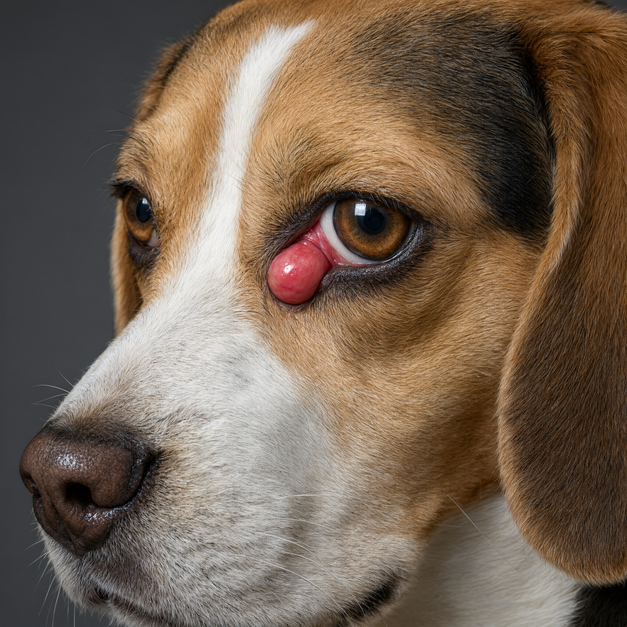
The classic “cherry” is a prolapsed tear gland of the third eyelid — not a tumor in most young dogs.
Dogs have a third eyelid (nictitating membrane) in the inner corner of each eye. Attached to it is a tear gland that produces a large share of the watery tear film — often cited as up to about 30–50% depending on the source and individual dog.
When the connective tissue that holds that gland in place is weak, the gland can slip forward and become visible as a smooth pink or red mass. That is cherry eye (prolapsed nictitans gland).
According to Dr. Yawar Abbas, DVM, MPhil, the goal of treatment is to keep the gland working. Removing it permanently raises the long-term risk of dry eye (keratoconjunctivitis sicca), which can damage vision and comfort.
Not every red mass is cherry eye. Corneal ulcers, foreign bodies, inflammation, and rare tumors need different care. Photos help your vet, but an in-person exam is required.
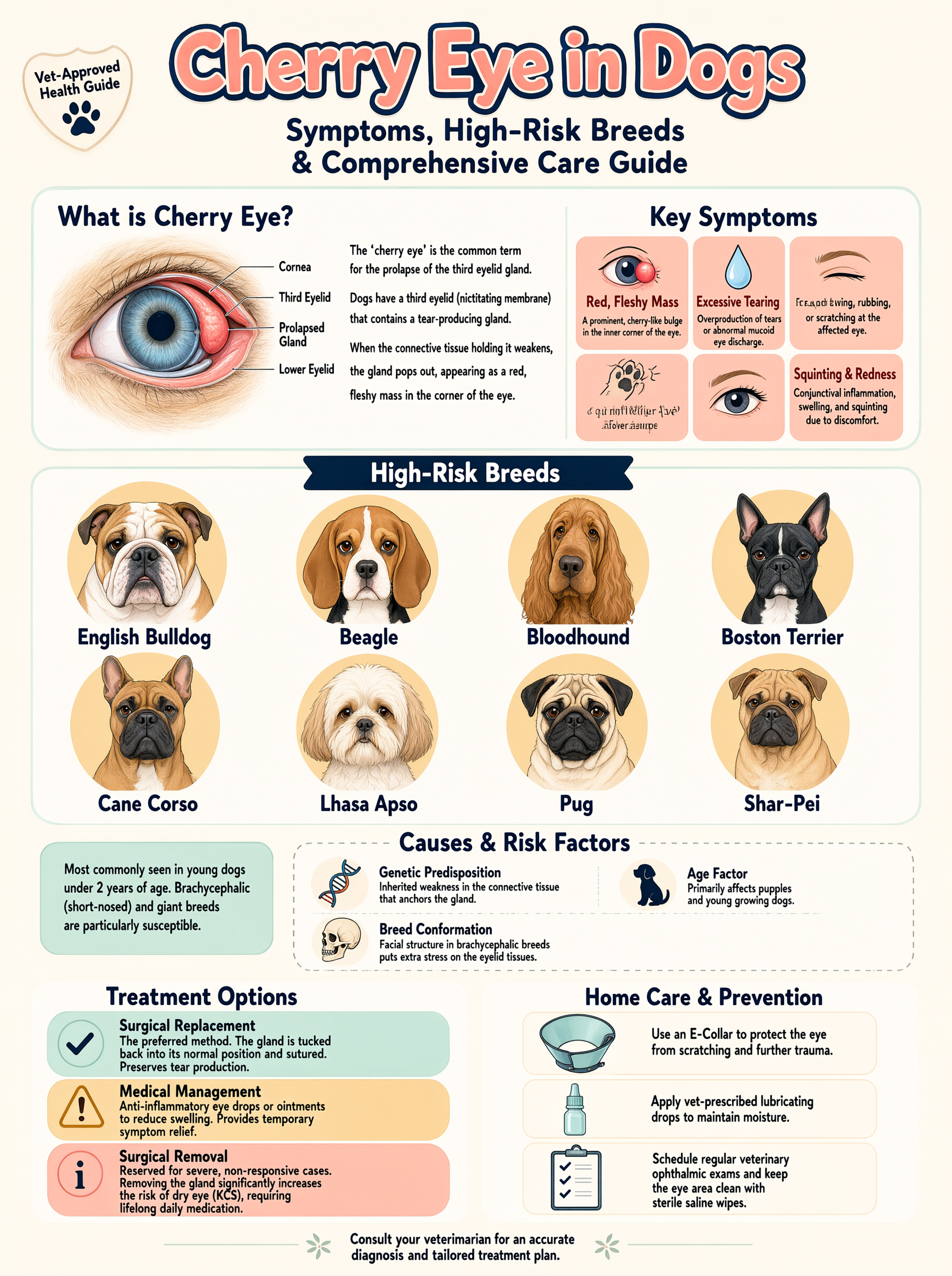
Young dogs in certain breeds show cherry eye most often — genetics and ligament strength matter.
Many dogs are not obviously painful at the start. Owners often ask “does cherry eye hurt” or “is cherry eye painful for dogs.” The prolapsed gland itself is usually more irritating than sharply painful, but the exposed tissue can dry, swell, and become inflamed. Rubbing raises the risk of corneal scratches.
Seek care sooner if the eye is held closed, discharge turns thick or colored, the cornea looks cloudy, or your dog seems depressed or stops eating.
The main driver is thought to be weak fibrous attachments of the third eyelid gland. Genetics and breed conformation play a large role. Trauma or chronic inflammation can contribute in some cases.
Breeds frequently listed at higher risk include:
Cherry eye is most often diagnosed in dogs under two years of age, including puppies.
Searches for “cherry eye in puppies” and “what is cherry eye in puppies” are common because the condition often appears early. Early veterinary evaluation helps protect tear production for the rest of the dog’s life. Waiting “to see if it goes away” usually delays the only durable fix in most cases: surgical replacement of the gland.
Diagnosis is largely visual, but a full eye exam may include:
| Approach | What it involves | Notes |
|---|---|---|
| Surgical replacement (preferred) | Gland is repositioned and secured (tacking or pocket/imbrication techniques) | Preserves tear production; standard of care |
| Medical management | Lubricants, anti-inflammatory drops when prescribed | May reduce swelling temporarily; rarely a permanent cure |
| Gland removal | Historical approach | Generally avoided because of dry-eye risk |
Your primary veterinarian may perform the surgery or refer to a veterinary ophthalmologist. Technique choice depends on anatomy, prior attempts, and surgeon preference. Recurrence can happen in a minority of dogs and may need a second procedure.
Online “cherry eye remedy for dogs” lists (oils, unguided massage, human redness drops) can delay care or injure the cornea. Lubrication and a cone may help comfort while you get to the clinic — they do not replace veterinary treatment.
Healthcare Consultations can help you organize questions about surgery timing, aftercare, and monitoring the other eye.
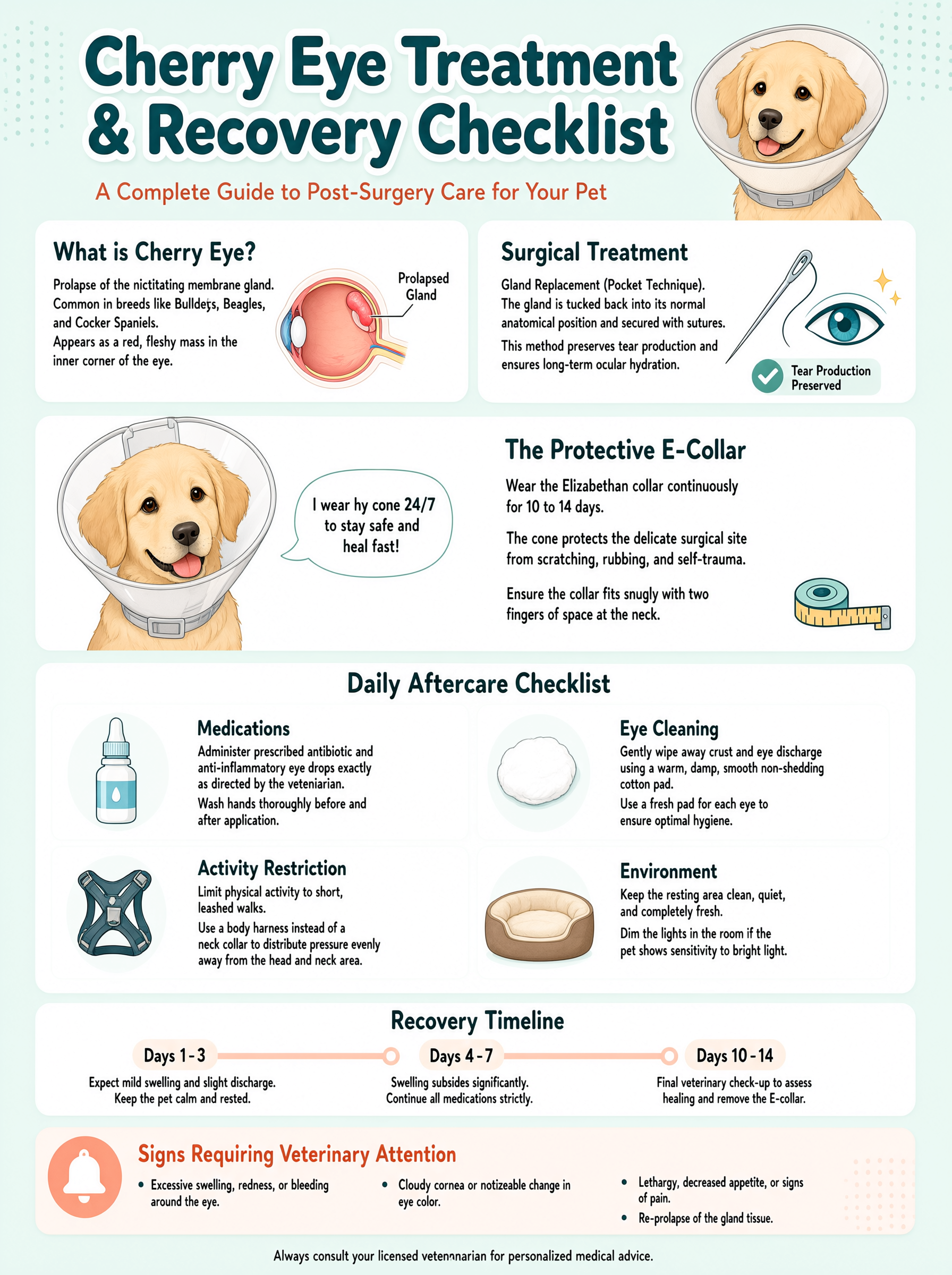
After surgery: cone, prescribed drops, and restricted play until your vet clears the eye.
Most dogs wear a cone and receive topical medications for about 10–14 days. Mild swelling is common early on. Contact your clinic promptly if the eye becomes suddenly painful, the cornea looks cloudy, or discharge worsens. Long-term outlook is good when the gland is preserved and tear production is monitored.
Will cherry eye go away on its own?
Usually not permanently. It may intermittently reduce then return. Most dogs need surgical replacement for a lasting result.
Is cherry eye an emergency?
Not typically, unless the eye is painful, ulcerated, or your dog is systemically unwell. Still schedule prompt veterinary care.
Can both eyes be affected?
Yes. Owners should watch the second eye even after one side is repaired.
How much does cherry eye surgery cost in the US?
Costs vary widely by region, clinic, and whether a specialist is involved. Ask your veterinarian for a written estimate that includes anesthesia, surgery, and recheck visits.
What is the best treatment for cherry eye in dogs?
Surgical replacement of the gland is the standard recommendation to protect tear production.
Cherry eye in dogs is common, especially in young dogs of predisposed breeds. It is usually more of a structural and tear-production problem than a sudden emergency, but delaying care can lead to dry eye and surface damage. Surgical replacement of the gland is the modern standard. Bookmark nextgenpetcare.com for more eye, puppy, and breed health guides.
Disclaimer: Educational only. Not a diagnosis or treatment plan. Seek hands-on veterinary care for any eye abnormality.